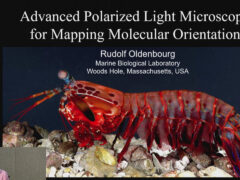
Advanced polarized light microscopy for mapping molecular orientation
- Rudolf Oldenbourg | Marine Biological Laboratory
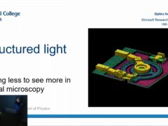
- Optics for the Cloud lecture series
Polarization is a basic property of light, but the human eye is not sensitive to it. Therefore, we don’t have an intuitive understanding of polarization and of optical phenomena that are based on it. And yet, polarized light plays an important role in nature and a suitably equipped polarized light microscope can be used to uniquely manipulate and analyze molecular order in materials, including living cells, tissues, and whole organisms.
I will discuss a modern version of the polarized light microscope called the LC-PolScope, which was developed at the MBL. Enhanced by liquid crystal devices, electronic imaging, and digital image processing techniques, the LC-PolScope reveals and measures the orientation of molecules in every resolved specimen point at once. In addition to birefringence and diattenuation, The LC-PolScope can also map the polarized fluorescence of fluorophores, including fluorescent proteins, that are used to label aligned structures like membranes and filamentous structures in living cells.
Recently we extended polarized light imaging to multi-view microscopes to overcome the restrictions inherent in traditional, single-view microscopes. With a traditional microscope, including the LC-PolScope, orientations are measured only in the plane perpendicular to the single viewing direction. Measuring the inclination angles of the optic axes of aligned bonds, particles, and fluorophores with respect to the focus plane remains a challenge. Multi-view microscopes, such as the light field microscope, can come to the rescue by recording specimen images along several viewing directions. This allows one to unambiguously measure the three dimensional orientation of molecules and their aggregates.
Series: Optics for the Cloud lecture series
-
Advanced polarized light microscopy for mapping molecular orientation
- Rudolf Oldenbourg
-
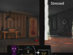
Modeling User Experience in Games: Lessons Learned
- Georgios Yannakakis
-
-